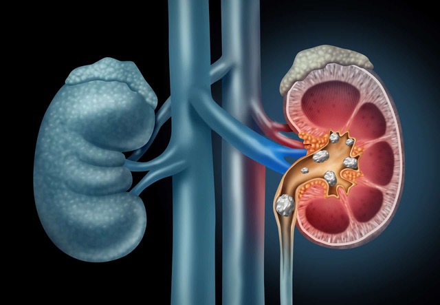
Urolithiasis is the third most common urological condition, following urinary tract infections and prostate disorders.
It refers to the formation of one or more stones in the urinary drainage system, which includes the renal pelvis and calyces, ureters, urinary bladder, and urethra. Urolithiasis is formed when there is an imbalance in kidney function—specifically, between water intake and the excretion of substances with low solubility. When this balance is disrupted, these substances accumulate into microscopic crystals, which gradually cluster together to form stones.

CAUSES
The causes of urolithiasis involve all factors that affect this delicate balance. Diet and metabolic disorders, the use of certain medications, genetic predisposition, urinary tract infections, and lifestyle habits such as fluid intake all play a significant role in stone formation.
TREATMENT
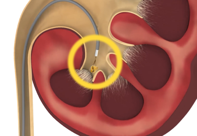
The management of urolithiasis is now, to a great extent, minimally invasive. A stone in the urinary tract — even if located high up in the kidney — can be accessed through the urethra, without the need for incisions. Using appropriate flexible instruments equipped with a light source and a camera at their tip (cystoscope and ureteroscope), the stone can be reached regardless of its location. Through the working channels of the endoscopes, laser fibers are introduced to fragment the stone, as well as flexible instruments (baskets) that can grasp and retrieve fragments and smaller stones.
The endoscopes themselves can either be used independently or passed through specialized working sheaths, which allow for improved irrigation of the operative field, optimal visibility, and the suctioning of smaller fragments.
Before the stone fragmentation procedure (lithotripsy), it may be necessary to place a flexible, self-retaining ureteral stent (pigtail) to relieve obstruction and alleviate symptoms Similarly, at the end of the procedure, a pigtail may be left in place for a few days, along with a bladder catheter if needed.

The development of increasingly thinner endoscopes that combine large working channels, excellent visualization, and outstanding flexibility and maneuverability allows for optimal effectiveness with minimal invasiveness and reduced risk of complications.
The procedure of accessing and fragmenting urinary stones through the urethra is called RIRS (Retrograde Intrarenal Surgery).
The use of such an endoscope, in combination with the powerful High-Power Holmium Laser MultiPulse HoPLUS by Jena Surgical, enables the endoscopic management of any stone with even greater safety, while minimizing operative time and the risk of residual stone fragments.
RIRS ADVANTAGES:
-
Minimally invasive, endoscopic procedure
-
Excellent effectiveness
-
Very low complication rates
-
Hospitalization up to 24 hours
-
Rapid return to daily activities

The approach to kidney stones through a small skin incision is called PCNL (Percutaneous Nephrolithotomy). This method is often preferred in cases of a large stone burden or when endoscopic access is not feasible or effective.
The development of smaller-diameter instruments and, more importantly, the use of a powerful laser such as the High-Power Holmium Laser MultiPulse HoPLUS by Jena Surgical have significantly reduced the invasiveness and potential complications of the procedure, making it a valuable tool in the hands of specialized urologic surgeons.
Finally, the combination of RIRS and PCNL is known as ECIRS (Endoscopic Combined IntraRenal Surgery). This technique is performed by experienced surgical teams and allows for the simultaneous treatment of all urinary stones—regardless of size and location—both endoscopically via the urethra and percutaneously through a small skin incision. This advanced technique fully utilizes modern technology and is based on the combined use of state-of-the-art, slim endoscopes used in RIRS along with a high-powered laser, ensuring faster and more effective management of the stone burden.
MULTIPULSE HOPLUS AVAILABILITY:
- Bioclinic Athens
- IASO GENERAL CLINIC
- ΝΤΥΝΑΝ
- COSMOCLINIC

